
JOURNAL OF THE DENTAL ASSOCIATION OF THAILAND
1
Page : 268-287Title : Success Rate and Survival Rate of Autotransplantation in Teeth with Completed Root Formation: Case Report 12 Cases
Author(s) : Chamroen Leelamanotham
Keyword(s) : Tooth autotransplantation,Success rate,Survival rate,Completed root formation
Manuscript Type : Case Report
Page : 268-287
The objective of this study was to determined success rate and survival rate of autotransplantation in teeth with complete root formation. The sample comprised 12 patients with 13 donor teeth (2 canines, 2 premolars, 9 third molars). The mean age of the patients at the time of surgery was 32 years (range 17 years 7 months to 56 years 8 months). Observation period were carried out at 1 week, 1, 3, 6 months, 1 year and once a year thereafter.The mean age of observation period was 4 years 4 months (range 1 year 3 months to 8 years 8 months). The follow up were examined clinically and radiographically. Clinical examination assessed plaque accumulation, gingival inflammation, pocket depth, tooth mobility and percussion test. Radiographs were used to examined bone healing, lamina dura, alveolar crest, periodontal space and root resorption. The success rate was 61.5 % (8 of 13 teeth) and survival rate was 92.3 (12 of 13 teeth). Autotransplantation in teeth with complete root formation has been proved by several researches of high survival rate. Therefore, autogenous tooth transplantation is one of viable treatment options for non-restorable tooth replacement.
2
Page : 288-295Title : The Role of Laser Stimulation (Electro-acupuncture) in Alginate Impression Making for Patients with a Gag Reflex.
Author(s) : Sanmati Pol, Farhin Katge, Vamsi Krishna, Manohar Poojari, Pooja Balgi, Ashveeta shetty
Keyword(s) : Acupressure,gagging prevention index,gag reflex,gagging severity index,laser acupuncture
Manuscript Type : Original Article (บทวิทยาการ)
Page : 288-295
A pronounced gag reflex (GR) is a problem for the acceptance and delivery of dental treatment for children. Despite a range of management strategies, lasers represent a quantum leap forward in the treatment of pediatric dental patients. The aim of this study was to evaluate the effect of low level laser stimulation (electro-acupuncture) in controlling the gag reflex of patients requiring upper alginate impression. Forty-five patients participated in the study, and were divided into three groups (Groups A, B, and C) of fifteen patients in each group. GR assessment was estimated by using the Gagging Severity Index (GSI). Group A underwent a red-light soft magnetic field laser stimulation (electroacupuncture) on conception vessel 24 (CV 24) for one minute (min). Group B underwent a combination of laser acupuncture of CV 24 and acupressure on pericardium 6 (PC 6). Group C, formed the placebo group. During laser acupuncture and acupressure, a second impression was taken and the Gagging Prevention Index (GPI) was evaluated. Both the GSI and the GPI were recorded at three different stages of the dental impression making procedure, stage I- an empty impression tray, stage II- with a loaded tray, stage- III ability to keep the impression in the mouth until the alginate sets. Statistical analysis was done using the SPSS version 21 software (SPSS Inc., Chicago IL). A significant decrease in GPI values as compared to GSI values, was observed after the laser acupuncture in Group A and B (p<0.05). The average improvement between the GSI and the GPI scores before and during laser acupuncture and acupressure in Group B was 53.6 %, in Group A was 34.2 % and in Group C was 2.81 %. When the mean values of GSI and GPI scores of the empty tray, loaded tray, alginate set were compared among the three groups A, B, C before and during laser acupuncture and acupressure, the results were statistically significant (p<0.05). The study concluded that both techniques, laser acupuncture on acupuncture points CV 24 and the combination of laser acupuncture on acupuncture points CV 24 and acupressure point PC 6 were effective methods in controlling the
gag reflex.
gag reflex.
3
Page : 296-306Title : Effect of Different Mouthwashes on the Elastic Force of Elastomeric Chain
Author(s) : Kanlaya Insee, Kanittha Chinprasert, Nattida Vipaschiwin, Natthaporn Juntila
Keyword(s) : Mouthwash,Elastomeric chain,Force decay,Orthodontics
Manuscript Type : Original Article (บทวิทยาการ)
Page : 296-306
The objectives of this study were to compare the effect of different types of mouthwashes on the elastic force of elastomeric chains and to compare the effect of pH in different types of mouthwashes that were exposed to the elastomeric chains. A total of 210 elastomeric chain specimens was exposed to five different types of mouthwashes (Colgate® Plax, Listerine® Cool mint, Systema® Japanese cherry blossom, Fluocaril® Ortho 123 and Punjasri®) andtwo control groups (distilled water and acetic acid). The elastomeric chains were submerged in artificial saliva at 37°C and were exposed to the solution twice a day. Force measurements were performed at six-time intervals (Initial, Day 1, 7, 14, 21 and 28). The result showed that statistically significant difference was found between the initial (Day 0) and Day 1 in all groups. The highest percentage of force decay occurred during the first day. And the test group of mouthwashes that had the highest mean of force remain in Day 28 was Systema® Japanese cherry blossom, followed by Listerine® Cool mint, Colgate® Plax, Fluocaril® Ortho 123 and Punjasri® respectively. In conclusion, the mouthwash that had the highest force decay in elastomeric chains was Punjasri® with no significant correlation between pH of mouthwash and elastic force degradation.
4
Page : 307-320Title : Effect of Cements on Survival and Complication Rates of Crowns: 10-Year Clinical Evaluation
Author(s) : Morakot Piemjai, Noppawan Adunphichet
Keyword(s) : Survival rate,Complications,Resin cement,Crowns
Manuscript Type : Original Article (บทวิทยาการ)
Page : 307-320
Acid-base cements mostly create microleakage at tooth-restoration interface, while 4-META/MMA-TBB resin cement can provide microleakage-free interface. Microleakage has influence on marginal seal and retention of fixed dental prostheses which might affect their survival and complication rates. This retrospective study was conducted to explore the effect of luting cements on 5 to 10 years’ survival rates of single crowns and their complications such as caries associated with restorations, pulp necrosis and detachment. Two types of applied cement, acid-base cement (zinc phosphate, zinc polycarboxylate or glass-ionomer) and resin cement (4-META/ MMA-TBB) were evaluated in this study. One hundred and fifty nine patients treated with at least 1 single crown were recruited for examination, given a total of 583 teeth were included in this study. All crowns were performed by post-graduate students of Prosthodontic Department, Faculty of Dentistry, Chulalongkorn University during the year 2005 and 2010. Survival condition was determined by the absence of extracted teeth or renewal prostheses, while evaluated complications were caries associated with crown, pulp necrosis or crown detachment. Data was analyzed using Kaplan-Meier method followed by log-rank test to evaluate the differences of survival and complication rates between types of cement at significant level of 0.05. It was found that 5- and 10-year survival rates of acid-base cement were 92.2 % and 67.0 %, while those of 4-META/MMA-TBB cement were 95.5 % and 87.1 % respectively. The complication rate of caries associated with crown, pulp necrosis and crown detachment of acid-base cement were 12.5 %, 7.3 % and 4.4 %, whereas those of 4-META/MMA-TBB cement were 1.4 %, 2.4 % and 0.0 % respectively. There
were significant differences in survival rate (p=.023), caries (p=.001) and detachment complications (p=.008) between cement types. In conclusion, crowns fixed with 4-META/MMA-TBB cement that can provide microleakage-free tooth-prosthesis interface have longer-term function and less complication.
were significant differences in survival rate (p=.023), caries (p=.001) and detachment complications (p=.008) between cement types. In conclusion, crowns fixed with 4-META/MMA-TBB cement that can provide microleakage-free tooth-prosthesis interface have longer-term function and less complication.
5
Page : 321-330Title : Shear Bond Strength of Resin Cements to Human Dentin under Self-Curing Mode
Author(s) : Pannipa Khumtong, Chaiwat Maneenut
Keyword(s) : Resin cement,Self-curing mode,Shear bond strength
Manuscript Type : Original Article (บทวิทยาการ)
Page : 321-330
To measure the shear bond strength (SBS) of a dual-cured resin cement to human dentin under self-curing mode, and compare it with the SBS of two self-cured cements. The SBS of two self-cured resin cements (Super-Bond C&B, Sun Medical Co. Ltd, Kyoto, Japan (SB); C&B Cement, Bisco Inc., Schaumburg, IL, USA (CB)) and one dual-cured resin cement (NX3 Nexus, Kerr Corporation, Orange, CA, USA (NX)) were measured according to ISO 11405. All resin cements were used according to the manufacturers’ instructions to bond to the dentin of premolar teeth using a stainless-steel cylinder mold and allowed to self-cure. Shear bond strengths were measured after 1h and 24h storage in a humidified lightproof box at 37o C. Super-Bond C&B exhibited the highest bond strength at both 1h and 24h ((19.2 ± 2.8) MPa, (34.0 ± 5.1) MPa respectively) and the lowest was C&B Cement ((5.2 ± 1.1) MPa, (10.5 ± 2.8) respectively) (p≤0.05). The bond strength at 24h in all groups were significantly higher than at 1h. Resin cements with different chemical formulations yield significantly different bond strengths to human dentin. The self-cured acrylic resin cement showed the highest bond strength and should therefore be preferred in clinical situations where the photo-curing light cannot transmit through the restoration. Time after bonding increased the bonding performance of all resin cements.
6
Page : 331-342Title : Microtensile Bond Strength of Repaired Ceramic Using Resin Composite with Universal Adhesive System Compared to Conventional Bonding System In Vitro
Author(s) : Chisanu Lertthawinchira, Sirivimol Srisawasdi
Keyword(s) : Microtensile Bond Strength,Repaired Ceramic,Thermocycling,Universal Bonding
Manuscript Type : Original Article (บทวิทยาการ)
Page : 331-342
The effectiveness of 2 types of ceramic repaired using resin composite and a universal adhesive were compared to a conventional adhesive. Leucite-reinforced glass ceramic ingots (IPS Empress® Esthetic; “EE”; Ivoclar Vivadent, Germany) and lithium-disilicate glass ceramic ingots (IPS e.max® Press; “EM”; Ivoclar Vivadent, Germany) were fabricated into 8x8x4 mm ceramic blocks with a total number of 288. The ceramic surfaces were wet-polished with silicon carbide paper and then treated with 9.5 % hydrofluoric acid (Ultradent® Porcelain Etch; Ultradent, USA). Resin composite (FiltekTM Z350 XT, shade A4; 3M ESPE, USA) was built-up with 2 adhesive systems, one half (“U”) using universal dental adhesive (Single BondTM Universal; 3M ESPE, USA) and the other (“C”) using total etch dental adhesive (AdperTM ScotchbondTM Multipurpose Plus; 3M ESPE, USA) combined with ceramic primer (RelyxTM Ceramic Primer; 3M ESPE, USA). The specimens were stored in water at 37°C for 24 hours and then subjected to thermocycling for 10,000 cycles prior to a microtensile bond strength (μTBS) test. The specimens were then divided into a group of 36, for 8 groups, according to type of ceramic, adhesive system, and storage condition. Modes of failure were analyzed using a stereomicroscope (ML 9300; MEIJI, Japan). Three-way ANOVA and a Bonferroni post-hoc test were used to analyze the data (n = 36, α = 0.05). There was no significant difference between the aged and non-aged groups (p = 0.207). However, a Bonferroni post-hoc test revealed significant differences among all tested groups. The highest μTBS was recorded by the “EMC” group (36.310±13.12), while the lowest was found in the “EEU” group (22.020±7.94). The μTBS between the resin composite and ceramic repaired using a conventional adhesive system was higher compared with a universal adhesive system, especially in the lithium-disilicate type.
7
Page : 343-359Title : Distribution and Factors Influencing Turnover Intention of Dental Specialists in Thailand
Author(s) : Siriruk Nakornchai, Ploychat Ingsakulrungruang, Songvuth Tuongratanaphan
Keyword(s) : dental specialists,distribution,Thailand,turnover intention
Manuscript Type : Original Article (บทวิทยาการ)
Page : 343-359
At present, Thai dental specialists work in various specialties; providing specific dental services, nationwide. However, there are many factors influencing the retention and turnover intentions of dental specialists. Previous studies on the distribution of Thai dentists were mostly limited to general dentists or included both general dentists and specialists. The objectives of this study were to investigate geographic distribution and to explore factors influencing the turnover intentions of dental specialists. The study was a cross-sectional survey. Data collection was from the Royal College of Dental Surgeons of Thailand and questionnaires. The data were analyzed using descriptive statistics, chi-square and binary logistic regression analysis. In 2015, there were 1,441 dental specialists, in which 51.7 % were non-residency training specialists (NRT) and 48.3 % were residency training specialists (RT). The data revealed that 83.1 % of dental specialists worked in the public sector, while 16.9 % worked in the private sector. Most of NRT group worked at the Faculty of Dentistry, while most of RT group worked under the Ministry of Public Health. Regarding geographic distribution, 37.8 % worked in Bangkok with the rest scattered in various regions Out of the 1,297 questionnaires, 652 were returned and 605 were complete. Only 23.4 % expressed a turnover intention. There were more specialists in the RT than in the NRT group who wanted to move their working locations. While over half of the NRT wanted to relocate to work in the private sector, most of the RT wanted to relocate to the public sector. The variables, which was most related to turnover intentions, included hometown and current work in the public sector. In conclusion, dental specialist mostly worked in public sector, and only one-fourth of them expressed turnover intention. Dental specialists who not working in their hometowns showed their interests to move than those already working in their hometowns.
8
Page : 360-369Title : Salivary and Plaque Fluoride Level after MU Caries Preventive Program in Daycare Centers
Author(s) : Siriruk Nakornchai, Warinthorn Phranet, Rudee Surarit, Tippanart Vichayanrat
Keyword(s) : Daycare center,Fluoride,MU caries preventive program,Plaque, Saliva
Manuscript Type : Original Article (บทวิทยาการ)
Page : 360-369
This study aimed to investigate and compare fluoride levels in saliva and plaque between the MU caries preventive program and a standard program. A randomized controlled trial was conducted on 77 preschool children from five daycare centers in Pathum Thani, Thailand. Children were randomly arranged into 2 groups: 1) a control group was provided a standard program including oral examination, oral hygiene instruction, diet advice and a fluoride varnish application; 2) a treatment group was provided the MU caries preventive program, which added extra interventions, including Interim Therapeutic Restoration (ITR) and sealant on posterior teeth with glass-ionomer cement. Plaque and saliva samples were collected before and after the program implementation at 24 hours, 1 week, 1 and 3 months, respectively. Salivary fluoride level was measured by a fluoride electrode, while plaque fluoride level was analysed by micro-diffusion technique and using a fluoride electrode (Model 96-09 Orion). The difference of plaque and salivary fluoride levels between the two groups was analyzed by Repeated ANOVA and Mann-Whitney U test, respectively. The treatment group showed a significantly higher plaque fluoride level than the control group at 24 hours (p<0.001), 1 week (p=0.018), and 1 month. (p=0.022). However, no significant difference was observed between the two groups at 3 months (p=0.228). The salivary fluoride levels showed the same tendency. The treatment group showed significantly higher salivary fluoride levels than the control group at 24 hours (p<0.001), 1 week (p<0.001), and 1 month (p=0.028). However, no significant difference was observed between the two groups at 3 months (p=0.055). This study was concluded that the plaque and salivary fluoride levels of children in MU caries preventive program were significantly higher when compared with the standard program at 24 hours, 1 week and 1 month.
1
Page : 179-188Title : Alternative Approaches in Pediatric Dental Caries Management
Author(s) : Apa Juntavee
Keyword(s) : Carious lesion,Remineralization,Chemomechanical caries removal
Manuscript Type : Review Article (บทความปริทัศน์)
Page : 179-188
New approaches in pediatric dental caries management aims to manage initial carious lesion by promoting remineralization to prevent the extension of the disease. At the present time, dental treatment has been focused on contemporary technologies for remineralization therapies on enamel surface and drill-less dentistry. The objectives of this review article are to make the summary of advanced materials for clinical approaches to carious lesion as well as minimal intervention to cavitated carious lesion in order to preserve tooth structure by chemomechanical therapeutics. New approaches in pediatric dental caries management not only reduce pain and anxiety during dental treatment but also enhance child cooperation and positive attitudes leads to long term successful in oral health development for children worldwide
2
Page : 189-196Title : Class II Division 2 Malocclusion in Growing Patient
Author(s) : Chatchai Chatmahamongkol, Supanee Suntornlohanakul
Keyword(s) : Class II division 2 malocclusions,Definition,Etiology,Treatment modalities
Manuscript Type : Case Report
Page : 189-196
The characteristics of Class II division 2 malocclusions are retroclination of maxillary central incisors, deep overbite, short lower facial height and upper incisor be covered by lower lip than usual. Deep overbite can effect the masticatory system and also the existence of teeth and periodontal tissue in long term. The main goals of treatment in this specific malocclusion are deep overbite correction, upper central incisors inclination correction with upper occlusal plane adjustment, correction of relationship between upper incisor and the lip and normal oromuscular function promotion. The objectives of this article are to present the knowledge involving orthodontics treatment in growing patient with Class II division 2 malocclusions.
3
Page : 197-211Title : Root Canal Therapy and Intentional Replantation of The Maxillary Central Incisor with A Combined Periodontic and Endodontic Lesion from A Palatogingival Groove: A Case Report
Author(s) : Kassara Pattamapun, Nuntiporn Jiamjit, Attapon Saelo, Julaporn Krisanaprakornkit, Suttichai Krisanaprakornkit
Keyword(s) : Palatogingival groove,Periradicular lesion,Root canal therapy,Tooth replantation
Manuscript Type : Case Report
Page : 197-211
The objective of this case report was to introduce a treatment approach for the maxillary central incisor, diagnosed with a combined periodontic and endodontic lesion from a deep palatogingival groove that originated at a palatal fossa of the crown and ended at the root apex. The palatogingival groove is often found in both maxillary lateral and central incisors and is regarded as a local factor that promotes an accumulation of dental plaque which is difficult to remove and eventually contributes to localized periodontitis. Moreover, the groove’s depth and length influenced the severity of inflammation in periodontium and dental pulp. In this case report, the authors have proposed a novel treatment strategy for the affected tooth, i.e., root canal therapy and intentional replantation, using a new root canal filling material with good sealability, OrthoMTA, along with Biodentine™ to repair defective dentine within the palatogingival groove.
4
Page : 212-224Title : Effect of Desensitizing Toothpaste on Microleakage of A Universal Dental Adhesive
Author(s) : Anyaporn Teekakul, Sirivimol Srisawasdi
Keyword(s) : Bonding agent,Desensitizing toothpaste,Microleakage,Universal adhesive
Manuscript Type : Original Article (บทวิทยาการ)
Page : 212-224
This research aimed to study the effect of desensitizing toothpaste containing strontium acetate and toothpaste containing arginine on the effectiveness of two bonding agents in class V cavities restored with resin composite. Cavity preparations were performed on buccal root surface of 65 permanent premolar teeth. The teeth were divided into 6 groups consisting of the use Scotchbond™ Universal Adhesive after brushing with Sensodyne® Rapid Relief, Colgate® Sensitive Pro-Relief™ and no brushing, compared to teeth bonded with Scotchbond™ Multi-Purpose Adhesive after brushing with the same toothpastes and no brushing. All teeth were restored with resin composite. After thermocycling and immersion in silver nitrate solution, the teeth were sectioned and evaluated using 4-interval scores under a stereomicroscope. The results showed that leakage score of Scotchbond™ Multi-Purpose Adhesive groups were statistically significantly higher than that of Scotchbond™ Universal Adhesive groups. However, Scotchbond™ Universal Adhesive groups demonstrated significantly difference between each group. Meanwhile, for Scotchbond™ Multi-Purpose Adhesive groups, there was no statistical difference among toothpastes and no brushing. In this study, it was concluded that both toothpastes did not have any effect on microleakage of Scotchbond™ Multi-Purpose Adhesive, whereas Colgate® Sensitive Pro-Relief™ showed more microleakage than Sensodyne® Rapid Relief when using with Scotchbond™ Universal Adhesive.
5
Page : 225-235Title : Comparison of Shear Bond Strength, Water Sorption and Solubility of 3 Glass Ionomer Cements for Direct Bonding of Orthodontic Brackets in vitro
Author(s) : Pintu-on Chantarawaratit, Samornphun Sinthawornkul, Pasutha Thunyakitpisal, Nonglax Thunyakitpisal, Sirithan Jiemsirilers, Onusa Saravari
Keyword(s) : Glass Ionomer Cement,Resin-modified Glass Ionomer Cement,Orthodontic Bracket,Shear Bond Strength,Water Sorption,Solubility
Manuscript Type : Original Article (บทวิทยาการ)
Page : 225-235
The purpose of this study was to evaluate shear bond strength, water sorption and solubility of three brands of glass ionomer cement adhesives; RU-HBM1 (a resin-modified glass ionomer cement prototype), Hy-bond Glasionomer CX (GIC) and Fuji Ortho LC (RMGIC) as orthodontic adhesives. For shear bond strength test, thirty extracted human premolar teeth were divided into 3 groups (n=10) and bonded to stainless steel brackets. Shear bond strength was measured by a universal testing machine with a 1.0 mm/min crosshead speed. For the water sorption and solubility tests, six disc specimens were prepared for each group. Water sorption and solubility of the different adhesives were calculated by the weight of samples before and after immersion in artificial saliva and after desiccation. Data were analyzed by one-way ANOVA. The RU-HBM1 had significantly higher shear bond strength than Hy-bond Glasionomer CX but lower strength than Fuji Ortho LC (p<0.05). In addition, the RU-HBM1 had the lowest mean water sorption value and was the only orthodontic adhesive that had a positive mean solubility value. In conclusion, RU-HBM1 provided adequate shear bond strength for clinical orthodontic purposes and showed a lower water sorption parameter than commercial glass ionomer cements.
6
Page : 236-249Title : Effect of A Desensitizing Agent Containing Glutaraldehyde and HEMA on Microtensile Bond Strengths of Resin Composite to Dentin
Author(s) : Yossawadee Manapakdee, Chaiyasri Thunpithayakul
Keyword(s) : Desensitizing agent,Glutaraldehyde,Microtensile bond strength
Manuscript Type : Original Article (บทวิทยาการ)
Page : 236-249
การศึกษานี้มีวัตถุประสงค์เพื่อประเมินและเปรียบเทียบผลของสารลดภาวะเสียวฟันที่มีส่วนผสมของกลูตารัลดีไฮด์และไฮดรอกซีเอธิลเมธาครัยเลท (สารกลูมา) ต่อค่ากำลังแรงยึดแบบดึงระดับจุลภาคของเรซินคอมโพสิตต่อเนื้อฟันด้วยสารยึดติดระบบต่าง ๆ ทั้งภายหลังการยึดติดที่ 24 ชั่วโมง และภายหลังการจำลองการใช้งานด้วยการเปลี่ยนแปลงอุณหภูมิ โดยใช้ฟันกรามแท้ของมนุษย์ จำนวน 96 ซี่งนำมาตัด และขัดให้เรียบจนเผยเนื้อฟันด้านแก้มลึกจากผิวฟัน 1.5 มิลลิเมตร สุ่มแบ่งเป็น 2 กลุ่ม คือ กลุ่มที่ไม่ใช้และใช้สารกลูมาก่อนการบูรณะฟัน สุ่มแบ่งฟันในแต่ละกลุ่มเป็น 3 กลุ่ม ก่อนการบูรณะฟันด้วยเรซินคอมโพสิต (Premise, Kerr, USA) ร่วมกับสารยึดติดดังนี้ 1. Optibond® FL (Kerr, USA) หรือ OF 2. ClearfilTM SE Bond (Kuraray, Japan) หรือ CS 3. ScotchbondTM Universal Adhesive (3M ESPE, USA) หรือ SU นำฟันตัวอย่างมาตัดเป็นชิ้นงานให้มีลักษณะเป็นแท่งสี่เหลี่ยมมีขนาด 1×1×8 มม.3 แบ่งชิ้นงาน เป็น 2 กลุ่ม โดยกลุ่มแรกนำไปทดสอบกำลังแรงยึดแบบดึงระดับจุลภาคภายหลังจากแช่น้ำ 24 ชั่วโมง และอีกกลุ่มนำไปผ่านการจำลองการใช้งานด้วยการเปลี่ยนแปลงอุณหภูมิระหว่าง 5 และ 55 องศาเซลเซียส จำนวน 5,000 รอบ ก่อนการทดสอบกำลังแรงยึดแบบดึงระดับจุลภาค โดยใช้เครื่องทดสอบ สากลที่ความเร็วทดสอบ 1 มม.ต่อนาที นำข้อมูลมาวิเคราะห์ความแปรปรวนสองทางและเปรียบเทียบเชิงซ้อนชนิดทูกีย์ที่ระดับนัยสำคัญ p<0.05 ผลการศึกษาพบว่าค่ากำลังแรงยึดภายหลังการใช้สารกลูมามีค่าลดลงอย่างมีนัยสำคัญทางสถิติในสารยึดติดทุกระบบ โดยภายหลังการยึดติดที่ 24 ชั่วโมง พบว่า กลุ่มที่ไม่ใช้สารกลูมามีค่าแรงยึดดังนี้ OF=63.47±6.96 เมกะปาสคาล CS=64.84±7.85 เมกะปาสคาล และ SU=53.72±5.80 เมกะปาสคาล กลุ่มที่ใช้สารกลูมามีค่าแรงยึดดังนี้ OF=58.95±6.86 เมกะปาสคาล CS=47.26±5.48 เมกะปาสคาล และ SU=48.04±4.61 เมกะปาสคาล เมื่อผ่านการเปลี่ยนแปลงอุณหภูมิ พบว่ากลุ่มที่ไม่ใช้สารกลูมามีค่าแรงยึดดังนี้ OF=53.94±6.62 เมกะปาสคาล CS=48.76±6.99 เมกะ ปาสคาล และ SU=43.96±5.66 เมกะปาสคาล กลุ่มที่ใช้สารกลูมามีค่าแรงยึดดังนี้ OF=51.98±6.75 เมกะ ปาสคาล CS=40.27±4.83 เมกะปาสคาลและ SU=42.66±5.41 เมกะปาสคาล
สรุปว่าการใช้สารกลูมาทำให้ค่ากำลังแรงยึดของเรซินคอมโพสิตต่อเนื้อฟันมีค่าลดลง
สรุปว่าการใช้สารกลูมาทำให้ค่ากำลังแรงยึดของเรซินคอมโพสิตต่อเนื้อฟันมีค่าลดลง
7
Page : 250-259Title : Important Factors that Need to Be Concerned During Tooth Movement in Periodontitis Patients
Author(s) : Mutita Wongsuwanlert, Pornputthi Puttaravuttiporn, Chidchanok Leethanakul
Keyword(s) : Guided tissue regeneration,Multidisciplinary treatment,Orthodontic treatment,Periodontitis
Manuscript Type : Original Article (บทวิทยาการ)
Page : 250-259
Nowadays, there are increasing number of periodontitis patients seeking orthodontic treatment due to problems of malocclusion or unesthetics. There have been some concerns giving orthodontic treatment in periodontitis patients than normal patient because of the inflammation and reduction of periodontium. Therefore, the orthodontists should know about background of disease, relationship of malocclusion and periodontitis and limitation of orthodontic treatment. This article focus on how to prepare and maintain healthy periodontium before, during and after orthodontic treatment including periodontal surgery especially guided tissue regeneration. Moreover, this article introduces the interrelationship of multidisciplinary team such as orthodontist, periodontist and general dentist.
8
Page : 260-267Title : The Effect of Different EDTA-Irrigation Time on the Microtensile Bond Strength of Resin Sealers and Root Canal Dentine
Author(s) : Uraiwan Chokechanachaisakul, Sutt Pansawangwong
Keyword(s) : AH Plus,EDTA,MetaSEAL,Microtensile bond strength,Root canal sealer
Manuscript Type : Original Article (บทวิทยาการ)
Page : 260-267
This study aimed to determine how the duration of EDTA irrigation affects bond strength. The 160 extracted human premolars were decoronated and embedded in resin block. Root canals were prepared by using the NiTi rotary files and distilled water irrigation, and irrigated with 5 % NaOCl. In group 1, this was followed by irrigation with distilled water, while in groups 2-5, this was followed by irrigation with 17 % EDTA for 1, 3, 5, and 10 min, followed by distilled water. Two specimens of each group were used for scanning electron microscopic observation. The remaining specimens were divided into 2 groups—AH Plus and MetaSEAL (n = 15 each). The specimens were prepared for microtensile tests. The failure mode was identified, and the bond strength value was analysed using one-way ANOVA and Tukey’s HSD post-hoc test. The 10-min EDTA-treated specimens (group 5) showed greater microtensile bond strength than non-EDTA-treated specimens (group 1) (p < 0.001) in MetaSEAL group. The fractured surface showed mixed failure accounted for the majority of failures in all groups. In SEM observation, the NaOCl group showed a smear layer covering the dentine surface, but the EDTA groups showed an absence of smear layer and various depths of demineralized dentine and exposed collagen. In conclusion, the duration of EDTA irrigation affected on the microtensile bond strength of the methacrylate resin sealer and root dentine.
1
Page : 119-132Title : A Review on Current Treatment Modality of Mandibular Prognathism
Author(s) : Udom Thongudomporn, Khitparat Kamoltham
Keyword(s) : Prognathic mandible, Class III treatment, Stability
Manuscript Type : Review Article (บทความปริทัศน์)
Page : 119-132
Mandibular prognathism has been described as one of the most severe maxillofacial deformities. The etiology involves systemic disease, genetic influence or neuromuscular imbalance. Treatment modalities include growth modification, comprehensive orthodontic treatment and combined orthodontic-orthognathic surgery. The early treatment attempts to restrain the prognathic mandible with external force. Skeletal anchorage is also currently used in conjunction with orthopedic appliance. The camouflage treatment is done in more severe cases using various techniques and other adjunctive procedures such as the use of skeletal anchorage and induction of regional acceleratory phenomenon. Mandibular set back can be done in combination with other surgeries to eliminate prognathic jaw. The surgical first approach and the minimal pre-surgical orthodontics (MPO) technique have been popular lately, but careful case selection is necessary. The stability and several factors that contribute to unfavorable treatment outcome are reported.
2
Page : 133-142Title : Maxillomandibular Advancement in Treating Obstructive Sleep Apnea
Author(s) : Chidsanu Changsiripun, Pajima Thaitammayanon
Keyword(s) : Maxillomandibular advancement,Orthognathic surgery,Obstructive sleep apnea
Manuscript Type : Review Article (บทความปริทัศน์)
Page : 133-142
Currently, the number of obstructive sleep apnea (OSA) patients has been increasing. OSA can be life threatening if it is not treated. The clinician can perform either conservative or surgical treatment. Treatment choice depends on several factors, such as OSA severity, patient needs, and the advantages and limitations of each treatment. Maxillomandibular advancement (MMA) is considered to be the most effective surgical technique for treating OSA and has a high success rate. However, this approach has disadvantages, such as the associated risks and cost. This review article presents an overview of MMA, including its effect in treating OSA, facial profile changes, complications, and stability after MMA.
3
Page : 143-151Title : Demineralized Tooth Matrix Used as A Bone Graft in Ridge Preservation: A Case Report
Author(s) : Srisurang Suttapreyasri, Warisara Ouyyamwongs, Butsakorn Akarawatcharangura
Keyword(s) : Demineralized tooth matrix,bone substitute,ridge preservation
Manuscript Type : Case Report
Page : 143-151
Alveolar ridge resorption after tooth extraction is frequent, clinically significant and makes the placement of an implant-supported restoration difficult. Different types of bone substitutes such as xenograft, allograft and hydroxyapatite have been used for ridge preservation. Tooth is a hard tissue with similar organic and inorganic compositions to bone, and thus it could be used as a potential bone graft substitute. In this case report, an autologous demineralized tooth matrix (DTM) was used to preserve and augment the alveolar bone after tooth extraction, before dental implant installation. After 3.5 months, the bone core was trephined for histologic analysis and the dental implant was placed. During the healing period, neither infectious occurrence nor unexpected clinical symptoms were observed. DTM demonstrated good soft and hard tissue contour maintenance. At the time of implant installation, the socket was completely filled with osseous tissue. A histological examination showed new bone formation and resorption patterns of the DTM particles. The clinical and histological findings suggest that filling an extraction socket with DTM is a good alternative for implant site preparation. The results of this case report confirm the resorption of the DTM over time and the formation of quality new bone at the graft site.
4
Page : 152-162Title : Knowledge and Clinical Practice of Antithrombotic Therapy among Dentists in Chiang Mai, Thailand
Author(s) : Krit Leemasawat, Chaiporn Karaket, Arintaya Phrommintiku
Keyword(s) : Antiplatelet therapy,Antithrombotic therapy,Dental procedure,Survey,Warfarin
Manuscript Type : Original Article (บทวิทยาการ)
Page : 152-162
This study aimed to survey the knowledge and clinical practice of dentists regarding antithrombotic drug management before dental procedures. All dentists who worked in Chiang Mai, Thailand between February 2014 and December 2014 were invited to answer the questionnaires via mail. The survey items consisted of 20 questions focusing on knowledge and clinical practice regarding antiplatelet therapy, warfarin therapy and guideline recommendations. From 186 invitation mails, 100 dentists (53.8 %) returned the questionnaires. Seventy-two percent of the participants recognized clopidogrel, while only 20 % knew ticagrelor. Over 90 % of the participants did not know the optimal duration of dual antiplatelet therapy after stenting. Approximately half of the participants discontinued aspirin before dental procedures in their patients, and 66.7 % of them required at least 7 days of discontinuation. Three quarters of the participants suspended P2Y12 inhibitors before their procedures, and 87.0 % of them required at least 5 days of discontinuation. Warfarin was discontinued in 71.6 %, 89.4 % and 93.5 % of the patients undergoing low-, moderate- and high-risk procedures, respectively. Approximately half of the participants were willing to perform low-risk dental procedures at the INR level of 2.1-4.0, while most of them preferred to perform moderate- and high-risk dental procedures at INR level of ≤2.0 (60.9 % and 72.5 %, respectively). All the participants were aware of the guideline recommendations, but there was a discrepancy between their practice and guideline recommendations to which they referred. The knowledge of antithrombotic therapy among dentists in Chiang Mai, Thailand was limited. A high percentage of dentists practiced differently from guideline recommendations. Some educational interventions should be done to improve the quality and safety of the medical care.
5
Page : 163-169Title : In vitro Accuracy Assessment of Electronic Apex Locator: RSUpex
Author(s) : La-ongthong Vajrabhaya, Kemachart Wangpitukwong, Khuanchanok Laongnualpanich, Pitchayapa Aroonraj, Kanyanun Ramayasinpong, Thanabat Yiampanomkun, Pimtida Watcharapreechawong, Sani Boonyakul, Thawat Kaewgun, Suwanna Korsuwannawong
Keyword(s) : Alginate model,Electronic apex locators,Root ZX,RSUpex
Manuscript Type : Original Article (บทวิทยาการ)
Page : 163-169
Aim: To evaluate the accuracy of RSUpex, a newly developed electronic apex locator (EAL), by comparing it with a standard apex locator (Root ZX). Methodology: Forty single-root lower premolar human teeth with completed apex formation were embedded in alginate model, which simulated tooth-surrounding tissue. The working length (WL) of each tooth was determined by using both Root ZX and RSUpex. The actual working length of each tooth was determined under a microscope. Results: The working lengths determined by both electronic apex locators varied ±0.5 mm from the apical foramen, which were within the acceptable criteria. The WLs measured by Root ZX and RSUpex were 0.5 mm short of the apical foramen (AF) of 28 canals (70 %) and 22 canals (55 %) respectively. Eleven canals (27.5 %) and 17 canals (42.5.5 %) were beyond the AF respectively for Root ZX and RSUpex, while WLs of 1 canal (2.5 %) from both EALs were at the AF. The intra-class correlation cofficient of both devices was 0.988, which indicates excellent accuracy. Conclusion: The accuracy of RSUpex in working length determination is comparable with Root ZX. Further studies are needed to evaluate the success of RSUpex in clinical settings.
6
Page : 170-178Title : Effect of Ethylenediaminetetraacitic Acid and Citric Acid on the Microhardness of Root Dentin
Author(s) : Chinalai Piyachon, Siripan Sutthisuwan
Keyword(s) : Citric Acid,Dentin,Root Canal,Ethylenediaminetetraacitic acid,Hardness
Manuscript Type : Original Article (บทวิทยาการ)
Page : 170-178
The purpose of this study was to evaluate the effect of 17 % ethylenediaminetetraacitic acid (EDTA) at pH 7.21 and 10 % citric acid at pH 1.55 on the microhardness of root dentin. Thirty human mandibular premolars were split longitudinally and randomly divided into six groups. Specimens were treated as follows: Gr1: 17 % EDTA for 1 min, Gr2: 17 % EDTA for 3 min, Gr3: 17 % EDTA for 5 min, Gr4: 10 % citric acid for 1 min, Gr5: 10 % citric acid for 3 min and Gr6: 10 % citric acid for 5 min. Dentin microhardness was measured with a Knoop indenter under a 50-g load and a 15-seconds dwell time. Data were statistically analyzed using Paired sample t-test, One-way ANOVA and Independent samples t-test at 5 % significance level. The results indicated that both solutions significantly decreased the microhardness of root dentin when contacted at 1, 3 and 5 minutes. The reductions of the microhardness values of root dentins that were exposed to 10 % citric acid at 3 and 5 minutes were significantly higher than those of EDTA groups. Under the conditions of this study, 17 % EDTA at pH 7.21 could be safely used as an irrigating solution without a change of dentin surface microhardness when contact time is 1-5 minutes.
7
Page : 107-118Title : Surface Electromyographic Studies on Masticatory Muscle Activity Related to Orthodontics: A Review of Literature
Author(s) : Udom Thongudomporn, Pattra Sumonsiri
Keyword(s) : Surface electromyography,Masticatory muscle,Malocclusion/Facial morphology
Manuscript Type : Review Article (บทความปริทัศน์)
Page : 107-118
The understanding of masticatory muscle function and its relationship with craniofacial morphology is essential to the field of orthodontics. Electromyography (EMG) has been used to assess muscle function both qualitatively and quantitatively. Many studies attempted to relate masticatory muscle activity with facial form, but the results have been inconsistent. The influence of the masticatory muscles on orthodontic treatment, specifically vertical malocclusion correction, and stability is still controversial. The purpose of this article is to review the relationship among masticatory muscle function, facial morphology and malocclusion based on the electromyographic studies.
1
Page : 1-14Title : Air Quality in Dental Clinic
Author(s) : Ruchanee Ampornaramveth
Keyword(s) : Air quality,Dental clinic,Infection control,Ventilation
Manuscript Type : Review Article (บทความปริทัศน์)
Page : 1-14
Dental treatments utilize the instruments those generate aerosol comprise of blood and saliva which able to spread the microbial among dental staffs and also the patients. The quality assurance of the air in dental clinic is crucial in control of the infection. This articles will explain how dental aerosol generate during dental treatment, methods of air microbial sample collection, index of microbial air contamination as well as strategies to reduce or eliminate the aerosol. The proper management of air in dental clinic is not only provide the safe working environment for dental staffs but also provide safety for the dental patients.
2
Page : 15-26Title : Mechanobiological Responses in Orthodontic Tooth Movement
Author(s) : Natkrita Wongsupa, Chidchanok Leethanakul
Keyword(s) : Bone remodeling, Cytokines, Neurotransmitters, Orthodontic force, Periodontium
Manuscript Type : Review Article (บทความปริทัศน์)
Page : 15-26
Orthodontic tooth movement results from appropriate force application of both compression and
tension which affect the response and remodeling of periodontium tissue surrounding the teeth. The tissue remodeling process arises through the inflammatory pathway. The compression side brings about bone J DENT ASSOC THAI VOL.67 NO.1 JANUARY - MARCH 2017 16 Article in Press resorption, while the tension side supports bone formation. As a consequence of this, tooth movement occurs. Moreover, the role of various periodontal cells and mediators such as cytokines and even neurotransmitters also support such a response during force application. The objective of this review article is to present the mechanobiological responses of periodontal tissues and alveolar bone in orthodontic tooth movement.
tension which affect the response and remodeling of periodontium tissue surrounding the teeth. The tissue remodeling process arises through the inflammatory pathway. The compression side brings about bone J DENT ASSOC THAI VOL.67 NO.1 JANUARY - MARCH 2017 16 Article in Press resorption, while the tension side supports bone formation. As a consequence of this, tooth movement occurs. Moreover, the role of various periodontal cells and mediators such as cytokines and even neurotransmitters also support such a response during force application. The objective of this review article is to present the mechanobiological responses of periodontal tissues and alveolar bone in orthodontic tooth movement.
3
Page : 27-42Title : Restoring Multiple Tooth wear by Increased Vertical Dimension Using All Ceramic CAD-CAM Restoration and Direct Resin Composite Restoration: A Case Report
Author(s) : ชลญา บำรุงเรือน, Rasmee Jindarojnakul, Phanomporn Vanichanon, Sirivimol Srisawasdi
Keyword(s) : Rehabilitation,CAD-CAM,Vertical dimension,Resin composite
Manuscript Type : Case Report
Page : 27-42
Multiple tooth wear with limited interocclusal space is a challenge for dentist to manage. Clinical case report revealed examination and treatment planning in the patient with multiple tooth wear. The teeth needed to be restored, but there was insufficient space for the restorations. The treatment plan was to increase vertical dimension even though the patient had no loss of vertical dimension. Mandibular molars were restored with all ceramic CAD-CAM restorations. Mandibular premolars and anterior teeth, were restored using direct resin composite restorations. After 6-month follow-up, the restorations exhibited acceptable function and esthetics. Hence, dentition rehabilitation with increased vertical dimension using all ceramic CAD-CAM restorations and direct resin composite restorations, which provide acceptable esthetics and strength, would be a treatment option for multiple tooth wear.
4
Page : 43-54Title : Comparison of Microleakage between Encapsulated and Manually-mixed Glass Ionomer Restorative Materials
Author(s) : Onauma Angwaravong, Sukarnjanat Silapason, Pachara Gulgovit, Naruwan Ruadrew, Orapan Wiparattanapong, Patimaporn Pungchanchaikul, Namchai Sooksuntisakoonchai
Keyword(s) : Capsule,Glass ionomer,Microleakage,Primary teeth
Manuscript Type : Original Article (บทวิทยาการ)
Page : 43-54
The objectives of this in vitro study was to assess the microleakage of encapsulated versus hand-mixed glass ionomer restorations. Eighty-four extracted human primary molars were randomly allocated into 6 groups. Each sample was prepared for Class II slot cavity and then restored with six different materials: Fuji IX GP capsule®, Ketac™ Molar Aplicap™, RIVA SC capsule®, Fuji IX GP®, Ketac™ Molar, RIVA SC®. The procedure of each glass ionomer restoration followed manufacturer’s instructions. All teeth were thermocycling for 500 cycles between 5°C and 55°C. After thermocycling the whole surface of each tooth was coated with nail varnish except for one millimeter around the restoration. The teeth were immersed in 0.5 % methylene blue for 4 hours and then sectioned mesiodistally. The sections were analyzed for microleakage under X30 magnification of a stereomicroscope. The median of the percentage of dye penetration between encapsulated and hand-mixed glass ionomer restorations were analyzed by the Mann-Whitney U statistic at 0.05 a level. The result showed the microleakage of encapsulated groups different from those of hand-mixed groups. However, Ketac™ Molar Aplicap™ encapsulated group showed statistically significant less than Ketac™ Molar (p = 0.002). But there were no statistically significant of microleakage between Fuji IX GP capsule® and Fuji IX GP® (p = 0.773), and there were no statistically significant of microleakage between RIVA SC capsule® and RIVA SC® (p = 0.207). In conclusion, the microleakage of encapsulated Ketac™ Molar Aplicap™ group appeared to be less than handy-mixed Ketac™ Molar group
5
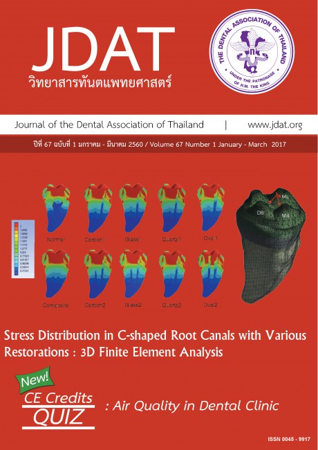
Page : 55-66Title : Stress Distribution in C-shaped Root Canals with Various Restorations : 3D Finite Element Analysis
Author(s) : Chinalai Piyachon, Nutta Pinyosopon, Jaruma Sakdee, Thiansiri Luangwilai
Keyword(s) : C-shaped root canal,Fiber post,Finite element analysis
Manuscript Type : Original Article (บทวิทยาการ)
Page : 55-66
The aim of this study was to investigate the stress distribution in C-shaped root canal restored with various restorations by 3D finite element analysis. Extracted permanent mandibular second molar was evaluated by micro-computed tomography. Ten 3-dimensional finite element analysis models were created and J DENT ASSOC THAI VOL.67 NO.1 JANUARY - MARCH 2017 56 Article in Press adapted by using the ANSYS computer software: normal tooth, tooth restored with resin composite, tooth restored with 1 and 2 carbon fiber posts, tooth restored with 1 and 2 glass fiber posts, tooth restored with 1 and 2 quartz fiber posts, tooth restored with 1 and 2 oval fiber posts. A 45-N load was applied using a 6-mm diameter of sphere on the occlusal surface. A tooth restored with resin composite core showed the highest amount of stress concentration which was higher than that of the normal tooth. Stress concentration of teeth restored with circular fiber posts was lower than that of those restored with oval fiber posts. The amount of stress in the teeth restored with 2-fiber posts were approximate to normal tooth and lower than that of those restored with 1-fiber post. Various restorations exhibited the high stress concentration at cervical area, especially on the buccal aspect. Under the condition of this study, it was concluded that C-shaped root canal
teeth restored with two glass fiber posts or two quartz fiber posts showed low stress concentrations when compared to the other types of restorations.
teeth restored with two glass fiber posts or two quartz fiber posts showed low stress concentrations when compared to the other types of restorations.
6
Page : 67-76Title : Effect of Silver Nanoparticles on the Compressive Strength of Acrylic Denture Base
Author(s) : Napapa Aimjirakul, Piyanart Ekworapoj
Keyword(s) : Acrylic denture base,Compressive strength,Silver nanoparticles
Manuscript Type : Original Article (บทวิทยาการ)
Page : 67-76
The purpose of this study was to investigate the effect of silver nanoparticles with different ratios on the compressive strength of the acrylic denture base. A total of eighty specimens (4 mm in diameter and 6 mm thickness) were fabricated from self-cured acrylic resin. Four groups (n = 20) were divided according to the concentration of silver nanoparticles incorporated to acrylic resin: 0, 1, 2 and 3 % w/w. All specimens were prepared for the compressive strength test using universal testing machine. The dispersion of silver nanoparticles was evaluated by Scanning Electron Microscope (SEM). Polymerization was analyzed by Fourier Transform Infrared Spectroscopy (FTIR). The result showed that the compressive strength of acrylic resin containing 2 % and 3 % of silver-nano was significantly higher than that of acrylic resin containing 1 % silver-nano and the control group (p < 0.05). SEM images confirmed the presence of silver-nano embedded to the polymer matrix. Nanoparticles are homogeneously dispersed over the specimen surface. In addition, polymerization of different ratios of silver nanoparticles was not different. This study showed that the concentrations of silver nanoparticles affect the compression strength of the acrylic resin. They are correlated: The higher the concentration of silver-nano, the greater the compressive strength. Moreover, the addition of silver nanoparticles
has no effect on the polymerization of acrylic resin
has no effect on the polymerization of acrylic resin
7
Page : 77-90Title : Physicochemical and Antimicrobial Properties of Silk Suture Soaked in Chlorhexidine Gluconate
Author(s) : Srisurang Suttapreyasri, Kemarajt Kemavongse, Phuntsho Choden, Kanvara Virojsakulchai, Prayoonsiri Kananuruck, Lalita Jeamkatanyoo, Vivara Watcharanuruk
Keyword(s) : Chlorhexidine gluconate,Antimicrobial,Physio-chemical,Suture material,Silk
Manuscript Type : Original Article (บทวิทยาการ)
Page : 77-90
The objectives of this study were to determine the physicochemical property, drug releasing pattern,
and antibacterial efficacy of silk suture soaked in chlorhexidine gluconate (CHX). The 3/0 black silk sutures
were immersed in 0.12 % or 0.20 % CHX for 24 hour. Then, the sutures were prepared by either dried at room
temperature (dry preparation) or stored in the CHX (wet preparation) and compared to the control untreated
silk suture in the aspect of the physicochemical property (tensile strength, surface morphology, porosity, and
existence of CHX), drug releasing pattern, and antimicrobial activity. Results showed that CHX-soaked silk
sutures exhibited greater diameter and average pore size compare to the control. There was no significant
difference of the tensile strength between CHX-soaked silk suture and control group. FTIR and EDX showed
CHX deposition on the surface and interstices of silk and burst release in first 24 hours before slow release
until the 7th day with no statistical significance between CHX groups. Antimicrobial efficacy was higher for wetthan
dry- preparation of CHX-soaked silk suture (p<0.05), while there was no significant difference between
the concentrations used. In conclusion, soaked silk suture with 0.12 % or 0.20 % CHX could accumulate and
release the CHX until the 7th day without compromising the physical properties of the sutures. The wet preparation
showed better antimicrobial property without the difference between the 0.12 % or 0.2 % concentration.
8
Page : 91-106Title : Effect of Various Polishing Techniques on Surface Roughness of CAD/CAM Ceramic Blocks
Author(s) : Palin Sappinan, Sirivimol Srisawasdi
Keyword(s) : CAD-CAM (Computer Aided Design-Computer Aided Manufacturing),Dental ceramic,Surface Roughness
Manuscript Type : Original Article (บทวิทยาการ)
Page : 91-106
The aim of this study was to compare the effect of various polishing techniques and overglazing on the surface roughness of Vita Mark™II blocks (70 specimens) IPS Empress CAD™blocks (70 specimens) and IPS e.max CAD™blocks (70 specimens). First, all specimens were ground with 50-micron diamond burs to simulate surface derived from Cerec milling system. The surface roughness was measured with a profilometer. Seventy blocks of each type of ceramic were divided into 7 groups (10 blocks each). Groups 1 and 2 were polished with Astropol™, groups 3 and 4 with SofLex™disc, groups 5 and 6 with OptraFine™system for 20 and 40 strokes respectively. Group 7 was overglazed. The mean values of average surface roughness (Ra) were compared using 3-way, 2-way and 1-way ANOVA, and Post Hoc Scheffe test (α=0.05). In Vita Mark™II groups, the lowest Ra was overglazed group. No significant difference in the mean surface roughness values was found between ceramic polished with OptraFine™system 20, 40 strokes, Astropol™ 40 strokes and SofLex™disc 40 strokes, which Ra were inferior to overglazed group. In IPS Empress CAD™groups, the lowest Ra was overglazed group. No significant difference in the mean surface roughness values was found between ceramic polished with OptraFine™system 20, 40 strokes, SofLex™disc 20, 40 strokes, which Ra were inferior to overglazed group. In IPS e.max CAD™group, the lowest Ra was overglazed group, no significant difference in the mean surface roughness values was found between ceramic polished with SofLex™disc 40 strokes and OptraFine™system 40 strokes, which Ra were inferior to overglazed group. From this study the smoothest surface for all ceramic was found in the overglazed group. The proper polishing method for Vita Mark™II was to polish with OptraFine™20, 40 strokes, SofLex™disc 40 strokes, Astropol™ 40 strokes, IPS Empress CAD™ with OptraFine™20, 40 strokes, SofLex™disc 20, 40 strokes, and IPS e.max CAD™ with SofLex™disc 40 strokes and OptraFine™system 40 strokes.
