JOURNAL OF THE DENTAL ASSOCIATION OF THAILAND

  • Issue 4 volume 66
  • Issue 3 volume 66
  • Issue 2 volume 66
  • Issue 1 volume 66
1
Page : 240-267

Title : Current Update in Human Saliva and Its Role in Oral and Systemic Health and Diseases

Author(s) : Ponlatham Chaiyarit

Keyword(s) : Diagnostics,Oral diseases,Saliva,Salivary gland,Systemic diseases

Manuscript Type : Review Article (บทความปริทัศน์)

Page : 240-267

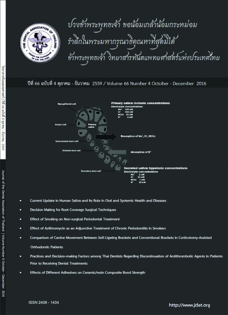
Human saliva is a complex secretion containing abundance of biomolecules derived from salivary glands, mucosal and periodontal tissues, and oral microorganisms. Formation of saliva is involved with coupling of the nerve-mediated reflex to glandular secretion of salivary fluid and proteins. A variety of molecules including peptides, proteins, glycoprotein, lipids, metabolites, RNA, and genomic DNA can be found in saliva. These salivary molecules are derived from both local and
systemic sources. Saliva has multifunctional roles in maintenance of oral health and supplies a variety of physiologically systemic needs including protection against tooth demineralization and microbial invasion, tissue lubrication, food perception, food digestion, and wound healing. Saliva can be an alternative source of other biofluids, because of the ease of obtainment, non-invasiveness and safety, and pleasantness of use. With an advanced-high throughput technology, a potential use of saliva as a diagnostic tool for oral and systemic diseases becomes essential for laboratory and clinical investigations with the aim of using saliva as a possible complementary examination with routinely diagnostic methods. The term “Salivaomics” was established recently to describe the information derived from studies in human saliva including: genomics and epigenomics, transcriptomics, proteomics, metabolomics and microbiomics. The aim of this review was to update the knowledge of human saliva regarding its role in oral and systemic health and diseases.

2
Page : 268-284

Title : Decision Making for Root Coverage Surgical Techniques

Author(s) : Kanoknadda Tavedhikul, Kajorn Kungsadalpipob

Keyword(s) : Recession,Root coverage techniques,Decision making for root coverage surgical techniques

Manuscript Type : Review Article (บทความปริทัศน์)

Page : 268-284

​Gingival recession has become patient’s esthetic concern. Several surgical techniques for root coverage have been proposed and modified to treat the recession defect and according to the review articles, the predictability and the success of root coverage varied among techniques. Decision in which technique to choose or how to manage this recession can be confusing. This articles focuses on indications, advantages and disadvantages of those techniques and provides the guideline of surgical techniques selection for successful treatment.

3
Page : 285-296

Title : Effect of Smoking on Non-surgical Periodontal Treatment

Author(s) : Navaporn Pornananrat

Keyword(s) : Non-surgical periodontal treatment,Non-smoking,Smoking

Manuscript Type : Original Article (บทวิทยาการ)

Page : 285-296

This study aims to compare periodontal status between smoking and non-smoking groups
after non-surgical periodontal treatment.
This cohort study consisted of 63 subjects (non-smoking 32 subjects and smoking 31 subjects) between November 2014 - November 2015. All subjects answered questionnaires and received periodontal examinations. Socioeconomic characteristics and smoking status were recorded by questionnaires at baseline. Probing depths were recorded at baseline, the 3-month follow-up and the 6-month follow-up. Data was analyzed by descriptive statistics and chi-square. Risk between smoking and non-smoking groups were compared by relative risk.
The results showed no difference between smoking and non-smoking groups with regard to socioeconomic characteristics except the difference among gender (p<0.01). At baseline periodontitis of smoking group was 32.79 %, non-smoking group was 27.86 %, the relative risk for periodontitis of smoking group 1.26 times greater than non-smoking group (95 % CI: 1.16 - 1.38). The effect of non-surgical periodontal therapy at the 6-month follow-up indicate periodontitis of smoking group was 9.55 %, non-smoking group was 3.43 %, the relative risk for periodontitis of smoking group 2.98
times greater than non-smoking group (95 % CI: 2.47 - 3.59).
The study indicates that smoking is harmful to periodontal status and outcome of non-surgical
periodontal treatment.

4
Page : 297-314

Title : Effect of Azithromycin as an Adjunctive Treatment of Chronic Periodontitis in Smokers

Author(s) : Putthiporn Chansuchai

Keyword(s) : one-visit full-mouth scaling and root planing,azithromycin,chronic periodontitis

Manuscript Type : Original Article (บทวิทยาการ)

Page : 297-314

There are numerous clinical studies in various antimicrobial agents improving the clinical outcome of chronic periodontitis therapy in smokers. Smokers with chronic periodontitis do not tend to respond to the periodontal therapy as favorable as non-smokers. The aim of this study was to compare the clinical results of one-visit full-mouth scaling and root planing with and without the use of azithromycin in smokers. 28 smokers with chronic periodontitis were divided equally into 2 groups, control group and test group. All clinical data were recorded; probing pocket depth, clinical attachment level, full mouth bleeding on probing and full mouth plaque index at 3 months and 6 months follow-up. The probing pocket depth and clinical attachment level were subdivided into 3 groups according to initial probing pocket depth as follows; 1-3, 4-6 and >7 mm, respectively. At 3 and 6 months, both therapies resulted in significant improvement when compared to baseline of probing pocket depth, clinical attachment level, full mouth bleeding on probing and full mouth plaque index. At 3 and 6 months, the clinical improvement in probing pocket depth and clinical attachment level in subgroup of initial pocket depth >7 mm. were significantly better in full-mouth periodontal therapy
with the use of azithromycin. In conclusion, the use of azithromycin as an adjunct to full-mouth periodontal therapy demonstrates favorable results in probing pocket depth reduction and clinical attachment level gain in smokers with advanced chronic periodontitis.

5
Page : 315-325

Title : Comparison of Canine Movement Between Self-Ligating Brackets and Conventional Brackets in Corticotomy-Assisted Orthodontic Patients

Author(s) : Bancha Samruajbenjakun, Thanawat Kiattawornwong, Narit Leepong

Keyword(s) : Canine retraction,Corticotomy,Self-ligating brackets

Manuscript Type : Original Article (บทวิทยาการ)

Page : 315-325

The purpose of this study was to compare the efficiency of maxillary canine movement when using self-ligating brackets with conventional brackets in corticotomy-assisted orthodontic patients. The study was performed on 18 sites in 9 patients (6 women, 3 men) with severe crowding who underwent first premolar extractions and corticotomy on maxillary canine areas. A conventional bracket was placed on one canine and a self-ligating bracket on the other side. Upper canines were retracted using elastomeric chains with 150 grams of force. After 3-months, impressions were taken every month and lateral cephalograms were taken at the beginning and the end of the experimental period. The results showed that in the self-ligating brackets group, the rate of maxillary canine movement was 1.62±0.27 mm/month and the canine distal tipping was 11.66°± 5.01°. Distopalatal rotation was 9.44°± 5.50°. In the conventional brackets group, the rate of maxillary canine movement was 1.37±0.39 mm/month. The canine distal tipping was 13.27°±5.71° and the distopalatal rotation was 9.22°± 6.07°. The difference in rates of maxillary canine movement, distal tipping and distopalatal rotation between the 2 groups were not statistically significant (p>0.05). Distal movement and rotation of the upper canines reinforced with corticotomy were similar when comparing conventional and self-ligating brackets. Rotation of the upper canines during sliding mechanics was minimized with conventional brackets

6
Page : 326-343

Title : Practices and Decision-making Factors among Thai Dentists Regarding Discontinuation of Antithrombotic Agents in Patients Prior to Receiving Dental Treatments

Author(s) : Wipaporn Chokchaivorakul, Niwat Phanpaisan, Bundarika Suwanawiboon, Chulaluk komoltri, Yongchai Nilanont

Keyword(s) : Anticoagulant,Antiplatelet,Antithrombotic,Dentist

Manuscript Type : Original Article (บทวิทยาการ)

Page : 326-343

The debate continues among dentists regarding whether to continue or discontinue antithrombotic medication in patients prior to dental procedures. As a result, protocols regarding antithrombotic vary widely. The objective of this study was to identify and analyze the behavior, knowledge and other factors that affect the decision among dentists to discontinue antithrombotic medication prior to dental treatment. This cross-sectional questionnaire-based study was conducted among licensed dentists practicing in Thailand. Ten thousand questionnaires were delivered to dentists who are members of the Dental Association of Thailand. The questionnaire consisted of questions about presurgical requirement and information relating to discontinuation of antithrombotic medication,
information about dental treatment given to patients, specialized field of dentistry and dentist work place. Of 10,000 distributed questionnaires, there were 1,838 respondents (18.38 %) with a mean age of 39.31±11.02 years and 32.2 % were men. One out of five dentists (21.7 %) indicated that they will not treat patients with concurrent use of antithrombotic medications. Most of those were university faculty members or dentists in private clinics; which were significantly higher as compared to dentists in other work places (p<0.05). Oral surgeons were significantly the least likely to interrupt antithrombotic medication (p<0.05). There were dentists who requested physician consultation before discontinuing antithrombotic medication prior to the dental procedures with a low bleeding risk, such as local infiltration anesthesia (17 %), restorative treatment (1.3 %), root canal treatment (13.9 %) and dentures (1.1 %). Misperceptions among dentist were also reported. Majority (64.8 %) of dentists consider an INR (International Normalized Ratio) value of lower than 2.0 to be safe for performing treatment. Almost 10 % of dentists reported assessing bleeding time value prior to performing a dental procedure. Identified factors that affect dentist decision to discontinue antithrombotic drugs include dental specialty, place of work, type of procedure and knowledge about patients receiving antithrombotic medication.

7
Page : 344-356

Title : Effects of Different Adhesives on Ceramic/Resin Composite Bond Strength

Author(s) : Awiruth Klaisiri, Nantawan Krajangta, Tool Sriamporn, Niyom Thamrongananskul

Keyword(s) : Adhesive,Feldspathic porcelain,Resin composite,Shear bond strength,Silane

Manuscript Type : Original Article (บทวิทยาการ)

Page : 344-356

The aim of this study was to evaluate the effect of three adhesive systems with and without silane application on feldspathic porcelain/resin composite bond strength. Three adhesive systems [Adper Scotchbond Multipurpose (SM), Adper Single Bond 2 (SB), Single Bond Universal (SU)] and one silane coupling agent [RelyX Ceramic Primer (Silane)] were used in this study. Sixty feldspathic porcelain disks 6 mm in diameter, 4 mm in thickness were prepared and embedded in dental gypsum. Specimens were polished with 400 and 600 grit silicon carbide paper, respectively. The samples were randomly divided into six groups (N=10 for each group) according to different treatments applied on feldspathic porcelain surfaces: Group1 (Silane-SM), Group2 (SM), Group3 (Silane-SB), Group4 (SB), Group5 (Silane-SU) and Group6 (SU). A silicone template (3 mm diameter, 2 mm thick) was placed on top of treated feldspathic porcelain surface. The resin composite was filled into the mold and then lightirradiated by light curing unit at a light intensity of 1000 mW/cm2 for 40 seconds. All bonded specimens were kept in distilled water at 37oC for 24 hours and they were subjected to shear bond strength
measurement using a universal testing machine at a crosshead speed of 0.5 mm/min. The data were statistically analyzed using one-way ANOVA and Tukey’s test (α=0.05). The shear bond strengths (MPa) of Group1 to Group 6 were 23.41±4.16, 12.66±0.91, 20.92±4.03, 10.80±1.43, 32.00±1.87, and 12.01±1.18 MPa, respectively. The shear bond strengths of Group1, Group3, and Group5 were significantly higher than those of Group2, Group4, and Group6 (p<0.05). Furthermore, Group5 showed the highest shear bond strength and was significantly higher than those of Group1 and Group3 (p<0.05).
In conclusion, the silane had a positive effect on shear bond strength between feldspathic porcelain and resin composite when used with the three adhesive systems. Silane application followed by adhesive application increased shear bond strength for all systems.

1
Page : 149-170

Title : Dentin Conditioning Agents for Glass-Ionomer Cement

Author(s) : Tool Sriamporn, Awiruth Klaisiri, Niyom Thamrongananskul

Keyword(s) : Dentin conditioning agent,Glass-ionomer cement,Polyacrylic acid

Manuscript Type : Review Article (บทความปริทัศน์)

Page : 149-170

The aim of this article was to review the properties of the dentin conditioning agents for glass-ionomer cement, for example, clean the tooth surface, modify or remove the smear layer, demineralize the tooth surface, increase the surface area and increase mechanical and/or chemical bond strength between the tooth surface and glass-ionomer cement. For many years, various tooth surface treatment agents have been available. These include citric acid, demineralizing solution, polyacrylic acid, ethylenediamine tetraacetic acid (EDTA), phosphoric acid and many others. Different study designs have been tested for these tooth surface treatment agents; for example, rinsing or not rinsing the agent with water after application or testing the effect of different concentrations of the agent. The most popular and widely used dentin conditioning agent for glass-ionomer cements is 10–30 % polyacrylic acid because of its similar composition to the liquid mixture of the glassionomer cement. Polyacrylic acid is a weak acid with high molecular weight that is capable of cleaning the tooth surface, modifying the smear layer, without inducing dentinal tubule opening when the proper concentration and application time is used, roughening the tooth surface and promoting the formation of chemical bond between hydroxyapatite and glass-ionomer cement. Nowadays, suppliers tend to sell their dentin conditioning products along with the glass-ionomer cements. Each of which has different chemical composition and different treatment method. Therefore, it is best to strictly follow the manufacturer’s instruction for the best result.

2
Page : 171-181

Title : Biodentine™ Partial Pulpotomy in Adult Permanent Teeth with Cariously-Exposed Pulp: Case Reports (up to 30 Months Follow-Up)

Author(s) : Patchanee Chuveera, Darunee Owittayakul

Keyword(s) : Biodentine™,Calcium silicate-based cement,Cariously exposed pulp,Partial pulpotomy,Mature permanent tooth

Manuscript Type : Case Report

Page : 171-181

A new calcium silicate-based cement, Biodentine™ (Septodont, Saint Maur des Fosses, France), has recently been introduced. With its biocompatibility and dentinal bridge-promoting properties comparable to those of the clinically well-accepted mineral trioxide aggregate (MTA), Biodentine™ could probably be an appropriate option for pulp capping procedures. However, clinical outcome studies on Biodentine™ are limited. Moreover, vital pulp therapy in cariously exposed permanent teeth is also controversial. These case reports describe clinical procedures and outcomes of partial pulpotomy using Biodentine™ as a pulp capping material in four cariously exposed adult permanent teeth in three patients aged 22 to 50 years. At 12 to 30 months follow-up, none of the patients experienced spontaneous pain, swelling or abscess formation. All teeth displayed a positive response to pulp testing and functioned normally. The radiographic evaluation revealed normal periapical areas.

3
Page : 182-192

Title : Spontaneous exfoliation of teeth and alveolar bone necrosis following herpes zoster Infection of trigeminal nerve: A case report and review of literature

Author(s) : Wannaporn Nilpanit

Keyword(s) : Herpes zoster,Spontaneous exfoliation of teeth,Alveolar bone necrosis

Manuscript Type : Case Report

Page : 182-192

Herpes zoster (HZ) is a disease caused by reactivation of varicella zoster virus (VZV). With the low immune condition, VZV was reactivated. It is characterized by multiple painful vesicles on skin that innervated by the affected nerve. The oral complications appear when the maxillary or mandibular divisions of the trigeminal nerve are involved. Oral complications are postherpitc neuralgia, devitalized teeth, resorption of root, and spontaneous exfoliation of teeth, osteomyelitis or alveolar bone necrosis. This paper presents a case of 54- year-old Thai male with underlying diabetic mellitus and hypertension presented with exposure of right mandibular alveolar bone necrosis and spontaneous exfoliation of right lower teeth after having vesicles at skin below right lower lip. Base on clinical features, a diagnosis of herpes zoster infection at right mandibular division of trigeminal nerve inducing alveolar bone necrosis with spontaneous exfoliation of teeth was given.

 
4
Page : 193-202

Title : A Comparison of Liners and Adhesive Systems in Preventing Coronal Dye Penetration in Root-filled Teeth Subjected to Functional Forces

Author(s) : Pattama Chailertvanitkul, Peerapong Kupradit, Poonyaphorn Thanyakoop, Pacharee Kulwattanaporn, Nuttapong Kantrong, Subin Puasiri, Paul Vincent Abbott

Keyword(s) : liners,adhesive system,dye penetration,functional forces

Manuscript Type : Original Article (บทวิทยาการ)

Page : 193-202

       Root-filled teeth must be restored with materials that prevent penetration of bacteria and fluids containing nutrients. Different liners materials and etching techniques are available for use with composite resins. The aim of this study was to compare dye penetration with two liners and two adhesive systems in access cavities of root-filled premolars whilst simultaneously subjected to functional loading. Extracted human premolars were accessed and root-filled. Teeth in groups I and II were lined Vitrebond while groups III and IV were lined with Ionosit. Groups I and III were etched primed with Optibond FL primer. Optibond FL adhesive was placed and filled with Premise composite resin. Groups II and IV had Optibond XLR primer, Optibond FL adhesive and the same composite. Specimens were then mounted and subjected to the equivalent of three months mastication whilst simultaneously immersed in India ink. Specimens were retrieved and sectioned. Dye penetration was measured and expressed as a ratio of the tooth length. Data were analysed by one-way ANOVA, followed by the Bonferroni test. The results showed that controls performed as designed. There was no statistically significant difference between the four experimental groups (p = 0.051). Neither Vitrebond nor Ionosit showed significant differences in preventing dye penetration (t-test, p = 0.663) but the total-etch system had significantly less dye penetration than the self-etch system (t-test, p = 0.007). In conclusions, there was no difference between the two liners in preventing dye penetration. The total-etch system was significantly better at preventing dye penetration than the self-etch system in ideal endodontic access cavities in root-filled premolar teeth.

5
Page : 203-212

Title : Tobacco Use and Cessation Training among Thai Dental Students - the Global Health Professional Student Survey, 2006 – 2011

Author(s) : Siriwan Pitayarangsarit, Wikul Visalseth, Surat Mongkolnchai-arunya, Jintana Peesing, Benjawan Danpradit, Thongchai Vachirarojpisan

Keyword(s) : Dental students,Smoking cessation,Thailand GHPSS,Training

Manuscript Type : Original Article (บทวิทยาการ)

Page : 203-212

This paper aimed to assess the changes on tobacco use, received cessation training and perceptions of policy banning smoking of dental students in Thailand by using the Global Health Professional Student Survey (GHPSS) conducted in 2006 and 2011. It was a census of 3rd year students from all 8 public dental schools in the country with 682 students in 2006 and 418 students in 2011 joining in the survey. The GHPSS results showed progresses in students’ learning experiences on smoking cessation. Their perception of being role model for smoking cessation increased from 98.0 % in 2006 to 98.3 % in 2011 and the need for cessation techniques increased from 80.9 % in 2006 to 91.9 % in 2011. Percentages of dental students reporting that they have learned cessation approaches to use with their patients increased from 14.1 % in 2006 to 32.7 % in 2011. In conclusion, the proportion of the 3rd year dental students that have learned the cessation approaches to use with their patients increased by double within 5 years. However, the contents of tobacco control training are different based on professors’ interest and there is a possible way to set a standard tobacco learning program among dental schools within the country.

6
Page : 213-227

Title : Competency in geriatric dentistry among Thammasat University dental graduates: Graduates and employers’ perceptions

Author(s) : Nattira Suksudaj, Jeerat Bhonsethalert, Thakolkorn Thakolkorn, Mongkol Puangpetch

Keyword(s) : Competence,Dental graduate,Geriatric dentistry

Manuscript Type : Original Article (บทวิทยาการ)

Page : 213-227

The aims of this study were to investigate self-perceived competences in geriatric dentistry of Thammasat University dental graduates, to investigate employers’ opinion on graduates’ competences and to compare the results from the two groups. The study population consisted of 289 students who graduated in 2000 - 2010 and 90 employers. The data were collected via selfadministered questionnaires which were sent to the graduates by post and online survey. The questionnaires included 28 items in six-point rating scale from 5 = very good to 1 = very bad and 0 = not applicable. The rating scores were later categorized based on the proportion of respondents who rated items as ‘good’ and ‘very good’, ie, ≥ 70 % = ‘excellent’, 60 - 69 % = ‘average’ and < 60 % = ‘need improvement’. The response rates of graduates and employers were 73 % and 31 %
respectively. Most graduates had more than five years working experience and few attended postgraduate training. Most graduates rated themselves as ‘need improvement’ in 75 % of the items whereas employers rated 46 % of items in the same category. Both graduates and employers agreed that graduates had excellent competences in identifying indications of using antibiotic prophylaxis and epinephrine in patients with cardiovascular diseases, managing clinical features in patients with diabetes mellitus and who are taking antiplatelet agents. In addition, both groups agreed on competences rated as ‘need improvement’ including identifying types of herbal/dietary supplements that affect anticoagulation function, taking history from patients/carers to perform assessment of activities of daily living and instrumental activities of daily living and evaluating nutritional deficiency from patients’ treatment record or history taking. In conclusion, employers agreed on most competences as perceived by graduates. However, graduates rated themselves as ‘need improvement’ in most items and this finding should be addressed in the development of effective geriatric dental education.

7
Page : 228-239

Title : The Comparative Study of Fracture Strength between CeltraTM Duo and IPS e.max® CAD

Author(s) : Nantawan Krajangta, Chayanit Angkananuwat, Lertrit Sarinnaphakorn

Keyword(s) : All-Ceramic restoration,CAD/CAM,Fractural strength,Zirconia-reinforced lithium silicate

Manuscript Type : Original Article (บทวิทยาการ)

Page : 228-239

This study aims to measure the fracture strength in molar region of new zirconia-reinforced lithium silicate (Celtra™ Duo, Dentsply) in comparison to lithium disilicate all-ceramic crowns (IPS e.max® CAD, Ivoclar Vivadent) made with computer-aided design/computer-aided manufacturing. Thirty standardized crown resin models were prepared with the same dimensions and randomly assigned to either Celtra™ Duo or IPS e.max® CAD group, fifteen each. Fractural load values were measured by a universal testing machine (Instron model 4464, USA) with a static load applied along the long axis of the specimen at a crosshead speed of 1 mm/min at room temperature until fracture. Fracture load values were recorded for each specimen and statistically analyzed by using unpaired t-test. The mean ± SD for the fracture load values were 1,696.67 ± 230.31 N and 1,569.87 ± 154.71 N for Celtra™ Duo and IPS e.max® CAD, respectively. There was no statistically differences of fracture strength between Celtra™ Duo and IPS e.max® CAD (p < 0.05).

1
Page : 72-80

Title : Work-Related Musculoskeletal Disorders Among Dentists

Author(s) : Nithimar Sermsuti-anuwat, Pornchai Sithisarankul

Keyword(s) : Dentist,Musculoskeletal disorder,Occupational health

Manuscript Type : Review Article (บทความปริทัศน์)

Page : 72-80

The health problems concerning muscle, tendons, ligaments, skeleton, cartilage and nerves which are inclusively termed “Work-related musculoskeletal disorders” are induced by normal performing dental work activities, could lead to temporary disorders or permanent disabilities. The potential occupational risk factors for musculoskeletal disorders are repetition, force, vibration, stress and posture. This article also introduces some chairside stretching exercises for strengthen the muscles and minimizing the risk of such disorders.

2
Page : 81-93

Title : Effect of Casein Phosphopeptide-Amorphous Calcium Phosphate (CPP-ACP) on Surface Microhardness of Human Enamel

Author(s) : Kornkamon Sukjit, Thunyarat Denkongphol, Pavana Jangvangsit, Rathagorn Putthacharoen

Keyword(s) : Casein Phosphopeptide-Amorphous Calcium Phosphate (CPP-ACP),Demineralization,Remineralization

Manuscript Type : Original Article (บทวิทยาการ)

Page : 81-93

The aim of this study was to evaluate the effect of Casein Phosphopeptide-Amorphous Calcium Phosphate (CPP-ACP) on surface microhardness of demineralized enamel surface of human tooth. Twenty-six extracted human maxillary premolars were kept in 0.1% thymol solution for 2 weeks after extraction. The tooth was cut vertically into 2 halves; buccal and lingual using diamond tooth cutting device. Fifty two specimens were randomly divided into 2 groups; control and experimental groups (N = 26 each). For demineralization, all specimens were stored in 0.1 M lactic acid pH 4.8 for 4 days. All speciemens were subjected to Vicker’s microhardness measurement (VHNC1 for control group and VHNE1 for experimental group). The specimens in control and experimental groups were stored in 0.1 M lactic acid pH 4.75 for 10 minutes twice a day and then in artificial saliva at 37ºC for 14 days, except that in experimental group, the specimens were applied with CPP-ACP tooth mousse for 3 minutes before storage in artificial saliva. After 14 days storage, the VHNC2, VHNE2 were measured and ΔVHNC and ΔVHNE which were the difference between VHNC1, VHNC2 and VHNE1, VHNE2 were calculated. In control group, All the VHN are VHNC1 = 242.07 kgf/mm2, VHNC2 = 201.69 kgf/mm2 and ΔVHNC = -40.37 kgf/mm2. In experimental group, all the VHN are VHNE1 = 245.31 kgf/mm2, VHNE2 = 259.62 kgf/mm2 and ΔVHNE = 14.31 kgf/mm2. ΔVHNC and ΔVHNE were tested by paired t-test (p = 0.05). Mean average of surface microharness of control group significantly differenced to study group (p < 0.001). The specimens treated with CPP-ACP showed significantly more microhardness value than those not treated (p < 0.001). It is concluded that CPP-ACP tooth mousse increased human enamel surface microhardness.

3
Page : 94-108

Title : Comparative Cytotoxicity and Biocompatibility of Two Aluminum Chloride Local Hemostatic Agents

Author(s) : Pintu-on Chantarawaratit, Pasutha Thunyakitpisal, Pravej Serichetaphongse, Watcharaporn Tasachan

Keyword(s) : Aluminum chloride,Biocompatibility,Cytotoxicity,Local hemostatic agent

Manuscript Type : Original Article (บทวิทยาการ)

Page : 94-108

The objective of this study was to evaluate the cytotoxicity and biocompatibility of two aluminum chloride containing local hemostatic agents, Racestyptine and a local hemostatic agent prototype, Dent-Chula100®. To determine cytotoxicity, primary human gingival and pulpal fibroblasts were treated with the local homeostatic agents at concentrations of 0.1, 1.0, 2.5, 5.0 or 10.0 % (v/v) for 15 minutes. The MTT assay was used, with untreated cells serving as control. The biocompatibility test was performed using the lower incisors of twelve 8-week old male Sprague Dawley rats.The free gingival margin of the lower central incisors was temporarily displaced for 15 minutes using retraction cords soaked with Racestyptine or Dent-Chula100®. Cords soaked with normal saline served as a control. Seven days after treatment, the lower jaws were dissected. The H&E stained tissue sections were histopathologically examined for four pathologic index scores; degree of sulcular epithelium damage, collagen fiber appearance and orientation, inflammation and vascular reaction. Two-way ANOVA and Kruskall-Wallis tests were performed for statistical analysis of the in vitro and in vivo studies, respectively (p < 0.05). Our data revealed that Dent-Chula100® at 5.0 % - 10.0 % significantly reduced gingival fibroblast cell viability compared with the control group (p < 0.05), while Racestyptine at 2.5 % - 10.0 % significantly decreased cell viability (p < 0.05). Both Dent-Chula100® and Racestyptine at 2.5 % significantly reduced pulpal fibroblast cell viability (p < 0.05). The histopathological data indicated that the periodontal tissues did not incur any significant damage after exposure to Dent-Chula100® or Racestyptine. In conclusion, the cytotoxicity of Dent-Chula100® and Racestyptine was dose-dependent. The in vivo study revealed that both Dent-Chula100® and Racestyptine were biocompatible with gingival tissues.

4
Page : 109-123

Title : Dimensional Accuracy and Surface Detail Reproduction of Polyvinylsiloxanether Impression Material Tested in Dry, Moist and Wet Conditions

Author(s) : Umaporn Vimonkittipong

Keyword(s) : Detail reproduction,Dimensional accuracy,Polyvinylsiloxanether

Manuscript Type : Original Article (บทวิทยาการ)

Page : 109-123

The purpose of this study was evaluated dimensional accuracy and surface detail reproduction of polyvinylsiloxanether impression (Identium®light) tested in dry, moist and wet conditions and compared with addition silicone (Panasil®contact plus X-light) and polyether (ImpregumTMPentaTMSoft). Ninety samples for each impression were replicated using stainless steel metal die according to American dental association (ADA) specification No. 19, 30 samples of each condition. Then, (1) dimensional accuracy of dry and moist samples was done by measuring the average length of the middle horizontal line of each impression by measuring microscope with 0.001 milimeters accuracy. Two-way analysis of variance (ANOVA) was used to compare the mean dimensional change of three impressions in dry and moist condition (α = 0.05) and studied about time factor by compared length of line between 1 hours and 24 hours by Paired t-test. (2) Surface detail reproduction of all above conditions was evaluated following the criteria specified in ADA standard for details reproduction: clear continuous replication at least 2 of 3 horizontal lines. Furthermore, this research also compared the amount of defects on impression. The change of length of dimension accuracy test after 24 hours compare with original model in each impression was signification and condition in dry and moist impression are not affected to dimensional change. ImpegumTMPentaTMSoft impression in moist condition was found the most dimensional change value at 24 hours and there is difference in dimensional accuracy after 24 hours and 1 hours in all types of impression (p < 0.05). In dry conditions, all impressions showed a definitive continuous horizontal line with no deformities. In moist conditions, Identium®light, Panasil®contact plus X-light and ImpegumTMPentaTMSoft showed a continuous line 86.7 %, 63.3 % and 100 % respectively. There were no difference in deformities in all three impression materials. In wet conditions, only the ImpegumTMPentaTMSoft impression passed the ADA standard, however many deformities were seen. This study shows dimensional change accuracy for three impression materials was within ADA standard (< 0.5 %) and the polyvinylsiloxanether impression showed excellent detail reproduction in dry condition but not as well as ImpegumTMPentaTMSoft in moist conditions.

5
Page : 124-132

Title : Effect of Herbal Toothpaste Containing Piper betle, Psidium guajava and Garcinia mangostana on Dental Plaque and Gingivitis

Author(s) : Supranee Benjasupattananan, Thera Rojassawasathien, Piyawan Jirayusakamol, Yanakorn Siriyeum, Jenjira Thechathaweewan, Wanlada Koheng, Chayanan Sirawuttipong

Keyword(s) : Gingivitis,Herbal toothpaste,Plaque control

Manuscript Type : Original Article (บทวิทยาการ)

Page : 124-132

The aim of this randomized controlled clinical study was to compare the efficacy of herbal toothpaste containing Piper betle, Psidium guajava and Garcinia mangostana with fluoridated toothpaste in the reduction of dental plaque and gingival inflammation. Fifty one healthy participants were randomly allocated to one of two groups, the control group (fluoridated toothpaste) and the test group (herbal toothpaste). The enclosed label toothpastes and new soft bristled toothbrushes were distributed to each participant according to their groups. All participants were instructed to brush their teeth with assigned toothpaste for 2 minutes, 2 times a day for 4 weeks. The plaque index and gingival index were examined and recorded on the first day (baseline), 14th day and 28th day (endpoint). The plaque index and gingival index scores were decreased statistically significant in both groups at the end of the study period (p < 0.05). However, these parameters were not statistically significant different between the groups.

6
Page : 133-148

Title : Effectiveness of Caries Infiltration and CPP-ACP Containing Paste on Color Change and Surface Hardness of Artificial White Spot Enamel Lesions

Author(s) : Vanthana Sattabanasuk, Patchareeya Pintanon, Danuchit Banomyong

Keyword(s) : Caries infiltration,Casein phosphopeptide-amorphous calcium phosphate (CPP-ACP),Color difference,Enamel caries,Surface hardness

Manuscript Type : Original Article (บทวิทยาการ)

Page : 133-148

Initial enamel caries is characterized as a whitish colored area with subsurface porosities that may have progressed to a more advanced lesion. Several approaches have been proposed to manage the non-cavitated enamel caries at different levels. This study aimed to determine the color improvement and surface hardness recovery of artificial white spot lesions after two conservative treatments - caries infiltration (Icon) and CPP-ACP containing paste (Tooth Mousse). Enamel carious lesions on extracted human premolars were created and underwent the two respective treatments following each manufacturer’s instructions. Groups with or without lesion formation, but no further treatments, were included as negative and positive controls. All specimens were subjected to the demineralization-remineralization pH cycles at 37 °C for 8 weeks. At designated time points, the surface microhardness of the lesions was assessed using Vickers diamond indenter and the difference in color of the lesions to the time before treatment was evaluated using a spectrophotometer. The lesions were also microscopically observed from both top and cross-sectional views using SEM. Parametric and non-parametric statistics were conducted to analyze all data at 95 % confidence level. Surface hardness of the lesions decreased significantly after artificial enamel caries formation. Following the caries infiltration treatment, hardness value recovered immediately and maintained throughout the period of pH cycles, even though not be comparable to that of the positive control. Difference in color of the lesions could be significantly detected after single treatment of the resin material. On the other hand, daily application of CPP-ACP containing paste did not regain the surface hardness of the lesions and the color change could not be significantly observed within 8 weeks of pH cycle. In conclusion, the caries infiltration is superior to the CPP-ACP treatment in color and surface hardness improvements of artificial white spot enamel lesions.

1
Page : 1-10

Title : Targeted Therapy of Oral Cancer: a Focus on COX-2 Inhibitors

Author(s) : Anak Iamaroon

Keyword(s) : COX-2 Inhibitors,Oral squamous cell carcinoma,Targeted therapy

Manuscript Type : Review Article (บทความปริทัศน์)

Page : 1-10

Oral squamous cell carcinoma (OSCC) is one of the major leading cancers worldwide. Generally, the incidence of OSCC remains constant globally but appears increasing in some parts of the world especially in the young group. Despite an easy access to early diagnosis and treatment, the mortality rate of patients with OSCC remains high due to delayed treatment and failure to control tumor recurrences and metastasis. The overall five-year survival rate for OSCC is considerably lower than other cancers and has not significantly changed during the last two decades. Recently, novel treatments, aiming to target specific molecules, aberrantly expressed during OSCC carcinogenesis, have been investigated and tested in clinical trials at several research settings. COX-2, in particular, is significantly overexpressed in OSCC and suggested to play a crucial role in OSCC carcinogenesis. This review will focus on the association between OSCC and COX-2 and the use of the COX-2 inhibitors as the targeted therapy for OSCC. This new approach in OSCC treatment offers hope that it may replace the conventional treatment modalities considered nonspecific and causing unwanted severe complications to patients.

2
Page : 11-19

Title : Lingual Osseous Choristoma: Report of Three Cases

Author(s) : Sompid Kintarak, Saipin Cheunchoksant, Thongchart Siripan, Patcharee Kumplanont

Keyword(s) : Choristoma,Osseous choristoma,Tongue choristoma

Manuscript Type : Case Report

Page : 11-19

Osseous choristoma is a rare entity. It represents the dense, mature, bony tissue in an abnormal location. We reported three cases of lingual osseous choristoma. All three lesions were presented in Thai females at or close to the foramen caecum. The lesions were smooth, round or ovoid and pedunculated in shape, and stony hard in consistency. The lesions were simply excised without local recurrence after five to nine years.
3
Page : 20-34

Title : Pulpal Response to Conventional Glass Ionomer Cement in Partial Pulpotomized Teeth in Dogs

Author(s) : Krittapas Tanakulwattana, Pairoj Linsuwanont, Chanin Kalpravidh

Keyword(s) : Conventional glass ionomer cement,Histopathological appearance,MTA,Partial pulpotomy,Pulp capping material

Manuscript Type : Original Article (บทวิทยาการ)

Page : 20-34

This research aimed to study the pulpal responses in dog’s teeth when conventional GIC (Ketac Fil PlusTM) was used as partial pulpotomy medicament in comparison to MTA (ProRoot MTATM). Cavity preparations were performed on 24 dog teeth until pulpal exposure was observed. The pulp was removed at the depth of 1 mm at the exposure sites. Teeth were randomly assigned into four groups; namely 1) conventional GIC 7 days 2) MTA 7 days 3) conventional GIC 70 days 4) MTA 70 days.
All pulpotomized teeth were restored with conventional GIC. At the periods of 7 and 70 days, the teeth were surgically extracted for histopathologic evaluation. Two teeth were excluded due to complete loss of restorative material, resulted in 22 remaining teeth for histopathologic evaluation. At 7 days period, MTA group had desirable pulpal response in terms of pulpal biocompatibility as in conventional GIC group. At 70 days period, MTA group demonstrated mild level of pulpal response while conventional GIC group showed various levels of pulpal response either mild, moderate levels or pulp necrosis. Within the limitations of the experiment, MTA showed more desirable pulpal response in terms of pulpal biocompatibility than conventional GIC.

4
Page : 35-43

Title : Management of Temporomandibular Disorder Patients by Ministry of Public Health General Dentists in the Central Thailand

Author(s) : Supang Chanton, Supranee Vicheinnet, Chanchai Hosanguan, Wantanee Mutirangura

Keyword(s) : Central Thailand,General dentists,Ministry of Public Health,Temporomandibular disorders,TMDs management

Manuscript Type : Original Article (บทวิทยาการ)

Page : 35-43

The aim of this study was to investigate the management of temporomandibular disorders (TMD) by Ministry of Public Health general dentists in the central Thailand. A questionnaire of TMDs management was developed and mailed to all Ministry of Public Health dentists in the central Thailand. The data were analyzed by descriptive statistics. Two hundred and seven general dentists
responded the questionnaire. The respondents comprised 57 (27.5 %) males and 150 (72.5 %) females, whose age ranged from 23 - 59 years (mean 31.7 years). One hundred and seventy one (82.6 %) respondents had encountered at least 1 TMD patient in the preceding year and 140 (67.6 %) managed TMD patients. The management provided by these 140 respondents were: providing patient education and self-care 140 (100.0 %), pharmacologic management 121 (86.4 %), physical therapy 129 (92.1 %), orthopedic appliance 66 (47.1 %), and occlusal treatment 52 (37.1 %). In conclusion, most general dentists had encountered and managed TMD patients. The dentists usually managed TMD patients with conservative and reversible treatment.

5
Page : 44-59

Title : Associations of Emotional Intelligence with Academic and Clinical Performance of Dental Undergraduates, Prince of Songkla University

Author(s) : Supawadee Naorungroj, Keerati Wongwiriyapokin, Jutharat Manuschai, Pichayaporn Ratti, Watcha Chinniyomwanich, Supharanan Horwongsakul

Keyword(s) : Academic achievement,Clinical performance,Dental education,Emotional Intelligence

Manuscript Type : Original Article (บทวิทยาการ)

Page : 44-59

This study aims to examine (i) levels of dental students’ emotional intelligence (EI); (ii) associations of EI with gender, year of study, and type of graduated high school; and (iii) associations of EI with average scores of knowledge, laboratory skills, and clinical performance. Of 122 5th and 6th year dental students, academic year 2014, Prince of Songkla University, 116 students had agreed to participate in the study and were included in analysis. Each dental student completed the Department of Mental Health’s Emotional Intelligence Questionnaire (DMH-EI). The questionnaire comprised of 52 self-assessment items, 9 domains identified as self-control, sympathy, responsibility, selfmotivation, problem-solving, relationship, self-esteem, life satisfaction, and happiness. Descriptive statistics were used to describe distribution of overall and sub EI scores. Statistical analyses were carried out using Independent-samples T-test, Pearson’s correlation and Chi-square to evaluate associations between EI with average scores as well as other factors. In this study, mean total EI score was 161.72 ± 13.78. Compared to norm-reference score, all sub EI score was in a normal range, except for self-control, which was above the norm. DMH-happiness scores of the 6th were higher than the 5th year dental students (p = 0.039). DMH-sympathy scores in female were higher than male (p = 0.046), while DMH-sympathy (p = 0.011) and DMH-responsibility (p = 0.012) scores of dental students graduated from public schools were higher than those graduated from private schools. DMHproblem- solving (r = 0.297; p < 0.01), DMH-self-esteem (r = 0.277; p < 0.01), and DMH-relationship
(r = 0.199; p < 0.05) scores were positively correlated with average clinical grades. This study indicates that some aspects of EI is associated with a noncognitive factor associated with dental students’ clinical performance.
 

6
Page : 60-69

Title : Serological Profiles of Hepatitis B Virus Infection among Pre-Clinical Dental Students at a University in Thailand

Author(s) : Piamkamon Vacharotayangul, Patrayu Taebunpakul, Surapoom Klawsirirojana

Keyword(s) : Hepatitis B,Professional education,Serology,Vaccination

Manuscript Type : Original Article (บทวิทยาการ)

Page : 60-69

       Thailand has implemented the HBV vaccination program for all newborns since 1992. The aim of this study was to investigate the serological profiles of HBV among pre-clinical dental students who were born before and in or after 1992 at a dental school, and to identify factors related to HBV serology in this population prior to their clinical practice. One hundred and eighty seven pre-clinical dental students participated in the study in 2011 - 2012. The information on seroprevalence of HBV infection, history of vaccination and immunization status, personal data, birthplace, work history, and risk factors related to HBV infection were assessed through a self-administered form. Blood collection was performed to investigate the level of HBsAg, anti-HBs antibody, and anti-HBc antibody. Descriptive statistics, prevalence ratio, with 95 % confidence interval, Chi-square test, and Fisher’s exact test were used for statistical analysis. There was one subject (0.53 %) with positive anti-HBc antibody, and no subject with positive HBsAg. There was no significant difference in anti-HBs antibody protectivity between the group of dental students who were born before and after 1992 when omitting the history of recent vaccination. History of receiving recent vaccination increased anti-HBs antibody level significantly. The results from this study show that dental students who were born in or after the integration of HBV vaccine into the national immunization program still need HBV screening test for HBV serological profile. Those who are not immunized to HBV should receive vaccination, either a full 3-dose or 1 stimulation dose prior to exposure of clinical work.